Home
/ Back Muscles Chart, Muscles Diagrams: Diagram of muscles and anatomy charts ... - The muscles of the lower back help stabilize, rotate, flex, and extend the spinal column, which is a bony tower of 24 vertebrae that gives the body structure and houses the spinal cord.the spinal.
Back Muscles Chart, Muscles Diagrams: Diagram of muscles and anatomy charts ... - The muscles of the lower back help stabilize, rotate, flex, and extend the spinal column, which is a bony tower of 24 vertebrae that gives the body structure and houses the spinal cord.the spinal.
Back Muscles Chart, Muscles Diagrams: Diagram of muscles and anatomy charts ... - The muscles of the lower back help stabilize, rotate, flex, and extend the spinal column, which is a bony tower of 24 vertebrae that gives the body structure and houses the spinal cord.the spinal.. For more anatomy content please follow us and visit our website: The fibres attach to the clavicle, acromion and the scapula spine. The achilles tendon in the strongest in the body. Symptoms of muscle pain include: An extremely strong tendon attached to the heel.
The most common causes of lower back pain are strain and problems with back structures. Muscles found in the superficial group include rhomboid major, rhomboid minor, levator scapulae, trapezius, latissimus dorsi. Certain back muscles extend to other areas, like the shoulders, upper arms, and thighs. Lie on your back with your knees bent and your feet flat on the floor (a). Three types of back muscles that help the spine function are extensors, flexors and obliques.
Female Muscles Diagram (With images) | Muscle diagram ... from i.pinimg.com The intermediate layer contains the erector spinae muscles, whose many functions include the extension and lateral flexion of the spine, head and neck. There are three different muscle groups found in the back: Brings hip away from body. Some of these muscles are quite large and cover broad areas. An extremely strong tendon attached to the heel. Artery) p.134 accessory nerve p. Related posts of muscles of the lower back and hip diagram muscle anatomy lab. Loss of control of the bowel or bladder and retention of urine may.
Muscle injuries of the lower back are commonly caused by an improper lift, lifting while twisting, or a sudden movement or fall, which may cause lower back pain.
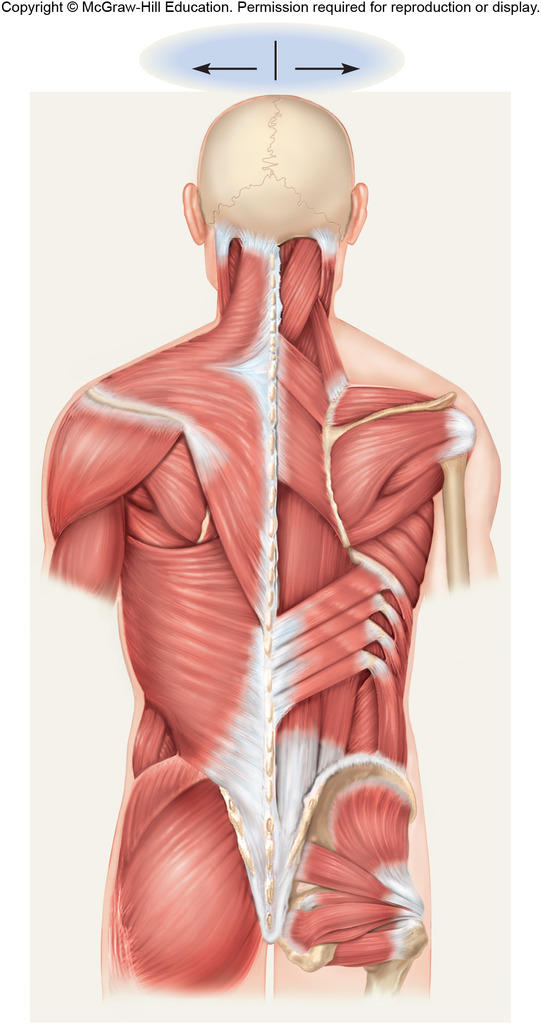
The superficial group, the deep group, and the intermediate group. There are a few warning signs, however, that may indicate serious spinal problems. The extensor muscles are attached to back of the spine and enable standing and lifting objects. The achilles tendon in the strongest in the body. Claim your free copy of the client back care guide today. For images of the muscle, click on each link under location. Chart of major posterior muscles. Related posts of muscles of the lower back and hip diagram muscle anatomy lab. The back consists of the spine, spinal cord, muscles, ligaments, and nerves. The teres major is a small, yet important muscle within the back. Loss of control of the bowel or bladder and retention of urine may. Our latest youtube film is ready to run. The rhomboid muscle is activated as you bring and squeeze your scapula or shoulder blades back and together.
To download your free copy click the link. Symptoms of muscle pain include: The superficial group, the deep group, and the intermediate group. Superficial, intermediate, deep and deepest layers.these muscles lie on each side of the vertebral column, deep to the thoracolumbar fascia they span the entire length of the vertebral column, extending from the cranium to the pelvis We are pleased to provide you with the picture named anatomy of back muscles diagram.we hope this picture anatomy of back muscles diagram can help you study and research.
Male Back Muscles Chart / Muscles German Names Chart ... from o.quizlet.com Brings leg back to and across body. 1) make midline incision along spines of vertebrae 2) extend from Lie on your back with your knees bent and your feet flat on the floor (a). It is the attachment site for the levator scapulae m. Strain commonly occurs with incorrect lifting of heavy. Your clients will thank you for it! Superficial, intermediate, deep and deepest layers.these muscles lie on each side of the vertebral column, deep to the thoracolumbar fascia they span the entire length of the vertebral column, extending from the cranium to the pelvis The teres major is a small, yet important muscle within the back.
The deltoid, teres major, teres minor, infraspinatus, supraspinatus (not shown) and subscapularis muscles (not shown) all extend from the scapula to the humerus and act on the shoulder joint.
Muscles found in the superficial group include rhomboid major, rhomboid minor, levator scapulae, trapezius, latissimus dorsi. It is the attachment site for the levator scapulae m. Raises and rotates arm in all directions. The achilles tendon in the strongest in the body. Brings hip away from body. They also protect the spinal column. These muscles include the large paired muscles in the lower back, called erector spinae, which help hold up the spine, and gluteal muscles. Just need a glimpse, leave your valuable advice let us know , and subscribe us! For more anatomy content please follow us and visit our website: Related posts of back muscles chart muscle anatomy stomach. The muscles on the back of the trunk help lower the arms and move the body forward and sideways. The vast majority of back problems improve on their own or with nonsurgical treatment. Muscle charts of the human body for your reference value these charts show the major superficial and deep muscles of the human body.
It is the attachment site for the levator scapulae m. Strain commonly occurs with incorrect lifting of heavy. The extensor muscles are attached to back of the spine and enable standing and lifting objects. It is an important site of muscle attachments for the intermediate layer of back muscles: It is the most superficial of all the back muscles.
Muscle Chart Back : Ab Muscles Abdominal Muscles Anatomy ... from images-na.ssl-images-amazon.com Just need a glimpse, leave your valuable advice let us know , and subscribe us! We are pleased to provide you with the picture named anatomy of back muscles diagram.we hope this picture anatomy of back muscles diagram can help you study and research. Using both hands, pull up one knee and press it to your chest (b). The angle of the scapula formed at the union of the superior and medial borders: Your clients will thank you for it! This procedure is one of the most powerful yet simple ways to treat muscle pain and discomfort. They extend and rotate the head and neck. Chart of major posterior muscles.
If you experience any of these symptoms, seek medical attention immediately.
The multifidus muscle keeps the back straight and stable. They extend and rotate the head and neck. The most common causes of lower back pain are strain and problems with back structures. Muscle anatomy stomach 12 photos of the muscle anatomy stomach female stomach muscle anatomy, human. A strain can be an injury to a tendon attachment from muscle to bone. These structures work together to support the body, enable a range of movements, and send messages from the. Deep back muscles diagram the superficial layer contains the splenius cervicis and splenius capitis muscles. Brings hip away from body. Three types of back muscles that help the spine function are extensors, flexors and obliques. The most common type of back pain is muscle pain—also called muscle strain or soft tissue strain. It is the attachment site for the levator scapulae m. Brings leg back to and across body. For images of the muscle, click on each link under location.

